


CARBOXYTHERAPY EFFECT ON THE CICATRIZATION OF CONTAMINATED SKIN WOUNDS OF RATS
Abstract
Objective: To investigate the effect of carboxytherapy in contaminated skin wound on the back of rats. Method: 15 Wistar rats weighting between 264g and 394 g (MD 313± IQD 18.50) were operated. In these animals a skin wound was produced their back with about 1.5 cm diameter and 0.5 cm depth. 24 hours after the animals were divided into two groups: control (n = 6) underwent puncture of the skin wound in its outer edge, subcutaneously, at 12 pm, 6 pm, 3pm and 9 pm; experiment (n = 9) submitted to the puncture wound in the same location as the control group and injected 2 ml of carbon dioxide in each location. This procedure was performed on the 1st, 2nd, 3rd and 4th and 5th postoperative days. Volumetry was held at 1, 4, 8 and 11 days. The animals were killed on the 11th day, then it was removed a skin fragment for histopathological examination. Results: The animals have evolved satisfactorily. There was reduction in the wounds of both groups. This was evidenced by the photographs of lesions and confirmed by volumetry in the same group (p<0.05). This phenomenon occurred in both groups. There were no differences between the macroscopic, microscopic and volumetric features (p> 0.05) of the wounds between the control group and the experimental group. Conclusion: carboxytherapy did not interfere with the cicatrization of contaminated open skin wound produced on the backs of rats.
Introduction
Wound treatment is described from the oldest times of human history. The first document that attests to the appearing of a wound in ancestors of men, Australopithecus africanus, has approximately 5 million years.1 This record shows a cranial lesion probably caused by a blunt object. The first caveman might have covered a wound with moss to avoid leaking a certain viscous material (hemostasis). The intentional surgical wound and trepanation were performed about 10.000 years ago and finger amputation in about at least 7.000 years. In the year of 3100 BC, in Mesopotamia it was discussed the issue of wounds. Egyptian papyrus of 2800 BC. to 1600 BC. attests to treatment of chronic lesions.2 The Egyptian would have been the first to use bandages on wound treatment. This people used animal fat and honey in the lesion, then covered it with a bandage of linen. The fat would create a physical barrier to contamination, honey had antibacterial function and linen would absorb secretion. Wound closing seems to have started in the 6th century, in ancient India, as was described in Susruta Samshita.3 The surgical instruments description suggests that intentional wounds were common and that lesions were used to facilitate healing.2 In this document was recommended debridement of loose and pulpy skin in burns.3 Hippocrates (470-360 BC) was in favor of primary intention cicatrization and the use of moist bandages. He was the author of “vis medicatriz naturae” aphorism, which means “the healing power of nature”. This included healing the wound. In the roman empire,
Cornelius Celsus described the acute signs of inflammation: flushing, tumor, warmth and pain. He said that acute wound would heal rapidly or spontaneously and that chronic wound would heal due to infection, mechanical irritation or inadequate circulation. Galen lived in the Greco-Roman period and preconized wine and suture to treat the wound. Posteriorly, for this purpose, it was used medication and local applications.4 In middle age, powders, ointments and cataplasms were used for the treatment of traumatic lesions. In Renaissance lived Ambroise Paré (1510-1590), father of Surgery, who condemned the cauterization and popularized hemostasis by ligation. He said “I tended to the wound, God healed it”, showing his humility. William Stewart Halsted (1852-1922), professor of surgery at Hospital John Hopkins, introduced surgery fundamental principles such as: not to pinch excessive tissue; use gloves during surgery; not use silk threads in infected wounds and perform rigorous hemostasis. In addition to these, it is assigned to Halsted other recommendations on wound treatment: gentleness, cleanliness, confrontation of the wound edges, good vascular supply, avoid tension, avoid dead space,5 which ultimately, favors tissue cicatrization. Contemporary medicine, despite all advances, is still challenged to treat many types of wounds.
In the treatment of open wounds, different types of medications have been used. In the experimental level it has been used the melipona honey,6 Aloe Vera,7 Laser,8 metronidazole,9 fibroblast growth factor,10 Passiflora edulis,11 allantoin,12 a combination of medium chain triglycerides, linoleic acid, soybean lecithin and vitamins A and E,13 Aroeira extract, aqueous extract of babassu,16 crude extract of jatropha gossypiifolia and propolis.18-20
Carboxytherapy has been used in treatment of many infirmities: stretch marks, cellulite, wrinkles, sagging, leg ulcers, peripheral artery disease with results still uncertain. Reports on carboxytherapy in contamined open skin wounds of rats were not found.
Considering that carboxytherapy has vasodilating effect, it was hypothesized that it might improve the healing of open skin wound. Therefore, it was performed this study aimed to evaluate the effect of carboxytheraphy in the cicatrization by secondary intention of contaminated skin wounds in the back of rats.
Method
After approval by the Animal Experimentation Ethics Committee of EMESCAM, 15 Wistar rats were studied, they were male, adults, with weight varying between 264g and 394g (M. 313 ± IQR 18.50). Each animal was punctured with one round shape wound in the dorsal region, in a way that the bottom margin of the wound would stay at the top of an imaginary line drawn between the ends of the scapulae, with 1.5 cm diameter, cutting the skin and subcutaneous tissue to the aponeurosis. The animals were randomly divided into 02 groups: group 1 (n = 9): carboxytherapy; Group 2: control (n = 06): puncture in the wound.
In the carboxytherapy group, it was performed a subcutaneous puncture in 4 different places with an insulin needle and 2ml of carbon dioxide was injected (12h, 6h, 3h and 9h) from the 1st to the 5th postoperative days (5 sessions). In the control group, the puncture was performed in the same places, with the same needle in the same days of carboxytherapy.
The rat’s dorsal wound was caused in the following sequence:
– Anesthesia with 5% ketamine hydrochloride was administered at a dose of 75 mg/kg/weight (Vetanarcol – Laboratory König – Inc – Argentina) and xylazine hydrochloride 2% at a dose of 8 mg/kg (Kensol – König Laboratory – Inc – Avellaneda- Argentina);
– Shaving of the dorsal region of 4cm2, with a shaving razor, performed after an imaginary line, cross section, from one ear to another, towards the dorsal-caudal;
– Local antisepsis with chlorhexidine at 10%, topical;
– Removal of skin fragment and subcutaneous cellular tissue, of round shape, with 1,5cm diameter and 0,5cm deep in the dorsal region;
– Wound hemostasis by local compression with gauze.
Photographs of the wound were taken for documentation.
The volume of the wound was evaluated at the 1st postoperative day by volumetry technique which was suggested by the research advisor (picture 1).21 This technique consists in the following steps: a – saline solution was dripped on the wound with micropipettes of 10 microliters and 100 microliters. Volumetry was re-evaluated on 4th day, 8th day and 11th day; b – The lesion was filled with saline solution; c – Verification on the micropipettes of volume necessary to fill the wound in microliters.
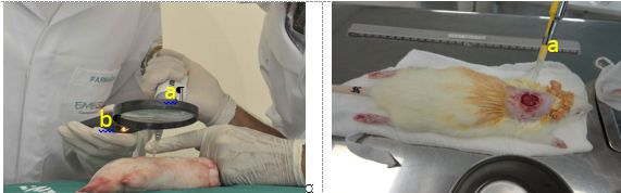
Figure 1 – Aspects of the technique of volumetry through micropipette (a) used to drip saline solution into the wound to calculate wound volume. A magnifying glass and lights (b) helped measure total filling of lesion and consequently its volume.
Source: the author.
Wound treatment (carboxytherapy) was performed with the machine Carbtek Advanced Dual Channel Plus (Manufacturer – DAF Produtos Hospitalares Ltda. Avenue Ibiúna, 86 – Vila Arucanduva 03507-010 São Paulo – SP – Brasil)
The animals were randomized (carboxytherapy group and punction group) as following:
– It was written the number 1 or 2 in a square chip with 2cm edge. 9 chips were numbered with number 1 (group 1) and 6 with the number 2 (group 2). Each chip was place in an opaque envelop, sealed. Then, one individual who wasn’t part of the study, scrambled and withdrew one chip which corresponded to the animal group (group 1 carboxytherapy and group 2 control). The researchers did not know which group the animal belonged.
After the treatment, the animals were returned to their cages and remained there with free diet and water. Analgesia of the animals was made by placing 200 mg of paracetamol in the water drinker and nubaim at 0.1ml/kg body weight subcutaneously.
In the day of surgery, carboxytherapy group, in the 1st, 4th, 8th and 11th days the wounds were photographed volumetry performed. In the 11th day it was removed a fragment of 3cm length by 3cm wide which included the wound in its central region, cutting the skin and subcutaneous of normal and wound areas. The animals were killed with Hypnol (3% sodium Pentobarbital – Syntec do Brasil Ltda) in dose of 120mg/kg.
The removed skin fragments were conserved in formalin 10% and then included in paraffin, submitted to transversal cuts of 4 μ with a microtome, and stained with hematoxylin-eosin (HE) to evaluate the histological changes.
Microscopy was performed by a binocular microscope pathologist, and the pathologist did not know to which group the animal belonged.
Statistic treatment
To calculate the median and standard deviation of the wound volume, descriptive statistics were used.
To compare the weight of the rats from the beginning to the end of the experiment, the Wilcoxon test was used.
To compare the volume of the wound between the different periods after surgery, in the same group, the Friedman test was used.
To compare the volume of the wound on the same day between the carboxytherapy and the control groups we used the Mann Whitney U test.
The tests were bicaudal, and p≤0.05 values were considered statistically significant.
Results
The wounds were produced without difficulties. Carboxytherapy was initiated 24 hours after wound production so it would be characterized as contaminated.
Animal weight had no significant difference between the beginning and the end of experiment in the carboxytherapy group of animals (median 313, interquartile range 24, median 330, interquartile range 46, p=0.594). However, in the animals of control group there was a significant weight increase (median 293, interquartile range 34.5, median 321.5, interquartile range 16.75, p=0.046).
It was observed similar closure of wounds in both groups, in visual macroscopic evaluation (Picture 2). There was a significant reduction in the wounds volume from the beginning to the end of the experiment both in carboxytherapy and control group (puncture) (p<0.05). The wound volume in control group in relation to carboxytherapy group had no significant differences in postoperative periods (table 1).
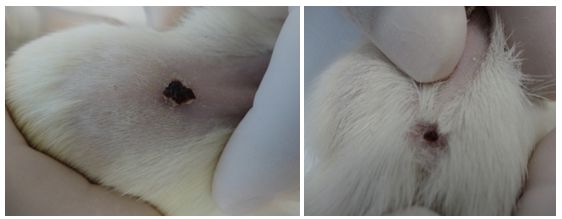
Figure 2: to observe the macroscopic aspect of the wound which was similar in the carboxytherapy group (Picture to the left) and control group (Picture to the right).
Source: the author.
Table 1 – Volumetry (in microliters) in the different postoperative periods between carboxytherapy and control groups.
| Postoperative Days | ||||
| Groups | 1st PO | Groups | 1st PO | Groups |
| M.D D.I.Q p | M.D D.I.Q p | M.D D.I.Q p | M.D D.I.Q p | |
| Carboxy | 163,33 ± 40,59 | 77,77 ± 22,79 | 16,00 ± 5,33 | 3,33 ± 0,86 |
| Control | 146,00 ± 18,41 0.60 | 71,66 ± 14,71 0.68 | 12,00 ± 3,63 0.06 | 2,33 ± 1,03 0.08 |
M – Median. IQR – Interquartile Range. PO – Postoperative
Mann Whitney test. p ≤0.05 – significant.
Source: the author.
Histological analysis showed in both groups, without difference, non-specific chronic inflammation, ulcerated with granulation tissue, edema and giant cell reaction of the foreign body type associated with surgical material. Surface presence of fibrin-leukocyte crust.
In the control group, histological sections stained with hematoxylin and eosin showed skin fragments exhibiting non-specific ulcerated chronic inflammation with tissue granulation and edema. There was no cicatrization difference between carboxytherapy and control group.
Discussion
Local treatment of open skin wounds is as ancient as the history of surgery.1-5 It involves cleaning, debridement, application of substances with possible cicatrizing effect and coverage or not of the lesion with gauze and tape. Despite the large number of locally applied substances7-20 that would have cicatrizing effect, there is still research for other methods because not always one method is effective for all wounds.
The weight of animals submitted to carboxytherapy had no significant variation from the beginning to the end of the experiment – 11th day (p>0.05). This is probably due to the stress followed by pain to which these animals were submitted. Besides, carboxytherapy causes reduction of subcutaneous fat volume. However, in the control group, not submitted to carboxytherapy, under lesser stressing situation, there was a significant weight gain from the beginning to the 11th postoperative day.
A challenge in the study of cicatrization is its evaluation. Although the ordinary methods of evaluation of cicatrization of an open skin wound are applied, none of them gives an accurate, quantitative sense, of the degree of closure of the lesion. Therefore, our proposal in this study was to use the volumetry of wounds21 giving a dimension of cicatrization in length, width and depth. The volumetry method of choice is to drip saline solution with the micropipette. Subsequent reviews of this method allow us to infer whether the wound is decreasing its volume (is cicatrizing) in a very objective way. It should be noted that volumetry is a recent method idealized by Paulo and Fiorot to evaluate cicatrization of an open wound. This is a safe method, because it evaluates the cicatrization that occurs from the periphery to the center of the wound, and from the bottom to the surface.
The idea of using carbon dioxide in the open skin wound was based on its vasodilator effect. Nevertheless, in this study, the subcutaneously injected carbon dioxide was not able to accelerate the cicatrization of open skin wounds of rats. There was no significant difference in the average volume of the wounds on the 1st, 4th, 8th and 11th days between carboxytherapy and the control group (p> 0.05). It is possible that the dose used was not optimal, as well as the carbon gas application time, or indeed carbon dioxide, despite its vasodilator effect did not alter scarring. This can be seen by analyzing the photos, which seems show no difference between the cicatrization of a wound from one group to another. The histopathological examination showed no scarring differences between one group and another, and neither did volumetry.
Conclusion
Carbon dioxide applied in the subcutaneous cellular tissue of a contaminated open wound of rats was not capable of accelerating cicatrization.
Acknowledgements
Thanks to Fundação de Amparo à Pesquisa do Espírito Santo (FAPES) for funding this research.
To the Instituto de Ações Solidárias do Espírito Santo for supporting this research.Authors
References
- Majno G. The healing Hand: Man and Wound in the Ancient World. Cambridge, MA: Harvard University Press: 1975) citado em Curr Probl Surg November 2007- pag 691-763.
- Robson MC, Steed DL, Franz MG. Wound healing: Biologic Features and Approaches to Maximize Healing Trajectories. Curr Prob Surg. 2001; 38(2): 71-140.
- Bhishagrama KKL. An English Translation of the Sushruta Samhita: Based on Original Sanskrit Text. Varanasi, India: Chowkhamba Sanskrit Series Office, 1963
- Paulo DNS, Loureiro ER. História da Cirurgia. In: Paulo DNS, Loureiro ER. Princípios de Cirurgia. 1ª ed. 1991. p.1-8.
- Romano LG, Nemetz AP. Cicatrização da ferida operatória. In: Nelson Fontana Margarido. Clin Bras Cirug. Vol. II. 1999. p.199-213.
- Alves DFS, Cabral Junior FC, Cabral PPAC, Oliveira Junior RM, Meneses do Rego AC, Medeiros AC. Efeitos da aplicação tópica do mel de Melípona subnitida em feridas infectadas de ratos. Rev Col Bras Cir (periódico na Internet). 2008; 35(3). Disponível em URL: http://www.scielo.br/scielo.php?script=sci_arttext&pid=S0100-69912008000300010
- Mendonça FA, Passarini Junior JR, Esquisatto MA, Mendonça JS, Franchini CC, Santos GM. Effects of the application of Aloe vera (L.) and microcurrent on the healing of wounds surgically induced in Wistar rats. Acta Cir Bras. 2009; 24(2): 150-5.
- Carvalho PTC, Mazzer N, Reis FA, Belchior ACG, Silva IS. Analysis of the influence of low-power HeNe laser on the healing of skin wounds in diabetic and non-diabetic rats. Acta Cir Bras. [ serial on the Internet]. 2006 May-June; 21(3). Available from URL: http://www.scielo.br/scielo.php?script=sci_arttext&pid=S0102-86502006000300010
- Trindade LCT, Biondo-Simões MLP, Sampaio CPP, Farias RE, Pierin RJ, Chomiski Netto. Avaliação do uso tópico do metronidazol no processo de cicatrização de feridas: um estudo experimental. Rev Col Bras Cir. 2010; 37(5): 358-62.
- Dantas Filho AM, Aguiar JLA, Rocha LRM, Azevedo IM, Ramalho E, Medeiros AC. Effects of the basic fibroblast growth factor and its anti-factor in the healing and collagen maturation of infected skin wound. Acta Cir Bras. 2007; 22(suppl 1): 64- 71.
- Garros IC, Campos ACL, Tâmbara EM, Tenório SB, Torres OJM, Agulham MA, et al. Extract from Passiflora edulis on the healing of open wounds in rats: morphometric and histological study. Acta Cir Bras. 2006; 21(suppl 3): 55-65.
- Araújo LU, Grabe-Guimarães A, Mosqueira VCF, Carneiro CM, Silva-Barcellos NMS. Profile of wound healing process induced by allantoin. Acta Cir Bras. [serial on the Internet] 2010 Sept-Oct; 25(5): 460-6. Available from URL: http://www.scielo.br/pdf/acb/v25n5/a14v25n5.pdf
- Magalhães MSF, Fechine FV, Macedo RN, Monteiro DLS, Oliveira CC, Brito GAC, et al. Effect of combination of medium chain triglycerides, linoleic acid, soy lecithin and vitamins A and E on wound healing in rats. Acta Cir Bras. 2008; 23(3): 262- 9.
- Branco Neto MLC, Ribas-Filho JM, Malafaia O, Oliveira Filho MA, Czeczko NG, Aoki S, et al. Evaluation of hydroalcoholic extract of Aroeira (Shinus Terebinthifolius Raddi) in the healing process of wound skin in rats. Acta Cir Bras (periódico na Internet). 2006; 21(supl 2). 17- 22. Disponível em URL: http://www.scielo.br/pdf/acb/v21s2/32158.pdf
- Amorim E, Matias JEF, Coelho JCU, Campos ACL, Stahke Jr HJ, Timi JRR, et al. Efeito do uso tópico do extrato aquoso de Orbignya pharelata (babaçu) na cicatrização de feridas cutâneas- estudo controlado em ratos. Acta Cir Bras [periódico na internet] 2006; 21(Suppl 2): 67-76. Disponível em URL: http://www.scielo.br/scielo.php?script=sci_arttext&pid=S0102-86502006000800011
- Martins NLP, Malafaia O, Ribas-Filho JM, Heibel M, Baldez RN, Vasconcelos PRL, et al. Análise comparativa da cicatrização da pele com o uso intraperitoneal de extrato aquoso de Orbignya pharelata (babaçu). Estudo controlado em ratos. Acta Cir Bras [periódico na internet] 2006; 21 (Supl 3): 66-75. Disponível em URL: http://www.scielo.br/pdf/acb/v21s3/10.pdf
- Santos MFS, Czeczko NG, Nassif PAN, Ribas-Filho JM, Alencar BLF, Malafaia O, et al. Avaliação do uso do extrato bruto de Jatropha gossypiifolia L. na cicatrização de feridas cutâneas em ratos. Acta Cir Bras [periódico na internet] 2006; 21 (Supl 3): 2-7. Disponível em URL: http://www.scielo.br/pdf/acb/v21s3/02.pdf
- Santos MJ, Vianna LAC, Gamba MA. Avaliação da eficácia da pomada de própolis em portadores de feridas crônicas/ the effect of própolis cream in healing chronic ulcers. Acta Paul Enferm. 2007; 20(2): 199-204.
- Segundo AS, Bosco AF, Maia D, Ribeiro RV, Aguiar EBH, Rocatto GEGD, et al. Influência do aloe vera e própolis na contração de feridas em dorso de ratos/ The Influence of Aloe vera and própolis in the contraction of wounds on dorsal área of rats. Periodontia. 2007; 17(1): 5-10.
- Peruchi CMS, Silva EB, Andrade RA, Franco SL, Ramalho LTO. Efecto del propóleos em la cicatrizacion de lesiones subcutâneas inducidas en el dorso de ratones: estudio histológico/ Effect of the própolis in induced subcutaneous wounds healing in mice: histologic study. Rev Fac Odontol Univ Chile. 2001; 19(2): 23-34.
- Paulo DN, Fiorot AW. A new proposal to evaluate the healing of open skin wound: volumetry. Acta Cir Bras. 2011; 26(6): 421-5
Authors
Danilo Nagib Salomão Paulo1; Claudia Cleto Pavan2; Lady Diana L. F de Almeida3
1 PhD from the Federal University of Minas Gerais – UFMG, Medical Doctor, Professor of Surgery Fundamentals of the Escola Superior de Ciências da Santa Casa de Misericórdia de Vitória – EMESCAM.
2,3 Medical Doctors graduated from the Escola Superior de Ciências da Santa Casa de Misericórdia de Vitória – EMESCAM.
