


SUBTOTAL SPLENECTOMY WITH INFERIOR POLE PRESERVATION (ESTPI)
Abstract
The spleen has important functions, within which there is the immune. Their withdrawal can result in the absence of these functions and hence infections occur, especially in children that can lead to death. Hence the importance of preserving the spleen or part of it. Of the conservative surgeries of the spleen (splenorraphy, segmental resections partial, autogenous splenic implant, subtotal splenectomy with preservation of the upper pole), the more recently described was subtotal splenectomy preserving the inferior pole (ESTPI). This operation, that constitutes in line of research of the first author, it aims to ensure the spleen functions.
Keywords
Spleen; General Surgery; Splenectomy
Introduction
Spleen is an organ of the mononuclear phagocyte system1 and for a long time it was considered to not be essential for life. Hence its removal due to trauma or other diseases does not cause greater harm to the patient.2-5 In 1952, King & Shumacker Jr found an association between spleen removal and the occurrence of sepsis in children.6 Since then, the risk of post-splenectomy infection have been recognized in children and adults, especially in the first two years after the surgery.7,8 Infectious complications after splenectomy were also observed in experimental animals.9-12 Changes have also been reported in humans’ lipid metabolism13-15 as well as in laboratory animals.16-21 Such changes can cause atherosclerosis.16 The spleen probably presents unknown functions that may be compromised by the organ’s removal. After observing spleen’s importance, its total or partial preservation started to be often treated through esplenorrhaphy,5 vascular occlusions,22-26 partial splenectomy27-29 and autogenous splenic implants.30-32 The most recently reported operation was the subtotal splenectomy with inferior pole preservation (ESTPI).33 This technique keeps blood supply by means of gastrosplenic ligament vessels that can be the existing vessels or the inferior polar artery vessel. The current study aims to review such technique.
Literature review
In 1999, the first author and collaborators published their pioneering work on the subject. They performed the ESTPI (Figure 1) in dogs to check the inferior pole viability.They used 24 dogs, weighing between 12 and 14 kg, divided into 2 groups: 1- ESTPI; 2- Spleen manipulation. Dog’s spleens and the remnants were removed on the 7th, 15th, 30th and 70th day, in group 1; and on the 15th and 60th day, in group 2, in order to study the macro and microscopic viability. The results showed that dog’s inferior pole spleen remained viable in 86.6% of cases, even with the splenic vessels ligature.33
In 2001, Paulo DNS and Lázaro da Silva,34 reviewed the literature on spleen and lipid metabolism and hypothesized that the spleen could interfere in lipid metabolism. Thereby, they performed an experiment in dogs to prove this hypothesis. They operated 38 dogs and their blood count and lipid profile were preoperatively performed and repeated on the 7th, 28th and 56th postoperative day. The animals were distributed into four groups: Group 1 (n=7) – spleen manipulation; Group 2 (n=9) – cranial hemi-splenectomy; Group 3 (n=9) – ESTPI; Group 4 (n=13) – total splenectomy (TS). Animals in Group 4 presented increased postoperatively total cholesterol, increased HDL fraction in the 7th and 28th day (p<0.05) and increased LDL in the 56th day (p<0.01). The VLDL fraction and triglycerides showed no significant differences between the preoperative and the postoperative period. Animals in groups 1 and 3 showed no significant changes and those in group 2 showed increased total cholesterol on the 7th PO day. Thus, the authors concluded that TS induces increased total cholesterol, HDL and LDL, and that hemi-splenectomy and ESTPI protects the animal from significant lipid changes.34
In 2005, Paulo ICAL et al35 conducted a study to evaluate the effect of total splenectomy, ligature of the principal splenic vessels and ESTPI on plasma lipid levels in rats fed with control diet or with diet supplemented with 2.5% cholesterol. They used 111 male Wistar rats weighing between 273 and 427g, approximately 12 weeks old. The animals were divided into five groups: Group 1 (n=20) – control, not operated; Group 2 (n=20) – spleen manipulation; Group 3 (n=31) – TS; Group 4 (n=20) – simultaneous ligature of the splenic artery and vein; Group 5 (n=20) – ESTPI. Plasma lipids were measured, and the rats were divided into two subgroups, according to the diet (subgroup A – control diet; subgroup B – supplemented with 2.5% cholesterol). All animals underwent a second blood test 90 days after the experiment. The animals submitted to total splenectomy, regardless of diet type and they showed significant increase (p<0.05) in total cholesterol, LDL, VLDL and triglycerides. HDL was significantly increased in rats fed with the control diet (p<0.05) and it was not significant in those fed with the supplemented diet (p>0.05). The animals underwent simultaneous ligation of splenic artery and vein and ESTPI and those fed with control diet showed no changes in plasma lipid levels, except for the HDL increase (p<0.05) observed in the subtotal splenectomy group. Animals fed with the supplemented diet (2.5% cholesterol) presented increase significant total cholesterol, LDL, and VLDL and triglycerides. Hence, the authors concluded that TS produces significant increase in total cholesterol, LDL and VLDL fractions and triglycerides, both in rats fed with the control diet and in those supplemented with 2.5% cholesterol. The increase was more significant in animals fed with the supplemented diet. The simultaneous ligation of splenic artery and vein and the ESTPI protects against changes in the plasma lipid levels found in rats submitted to TS and fed with the control diet or with 2.5% cholesterol diet 35 At that time, was held the first ESTPI in rats (Figure 2).
In 2006, Paulo DNS et al36 studied technical, morphological and functional aspects of the spleen inferior pole in rats. They operated 36 adult Wistar rats, divided into three groups: Group 1(n=10) – simulation; Group 2 (n=16) – TS; Group 3 (n = 10) – ESTPI. On the preoperative and on the 90th PO day, blood collection for cholesterol dosage, fractions and triglycerides was done. Next, the animals were deaths and their spleen and inferior pole were removed for macro and microscopic examination. Postoperatively, there were six deaths in the TS group; rats’ spleen and inferior pole presented to be macroscopically viable. The mean inferior pole weight was 35.84% ± 4.31. The animals in the group 1 and 3 showed no change in lipid profile between the pre and the postoperative. However, animals in the TS group showed significant increase in cholesterol and fractions. It was concluded that ESTPI was enforceable, that morphology of the inferior pole was apparently normal and that ESTPI prevented against lipid disorders. The authors noticed that, during the preservation of inferior pole supplied by gastrosplenic ligament vessels, the stump of the splenic artery inferior branch can be tapped where the gastrosplenic ligament does the anastomosis before entering into the spleen inferior pole.36
In 2007, Paulo ICAL et al37 observed the effects of hyperbaric oxygen therapy (HBO) (Figure 3) on 78 Wistar rats submitted or not to HBO. The animals were divided into three groups: Group 1 (n=20) – spleen manipulation; Group 2 (n=36) – autogenous splenic implant; Group 3 (n=22) – ESTPI. Each group was divided into two subgroups: a- not submitted to HBO (nsHBO); b- submitted to HBO (HBO). Then it was: Group 1, subgroup a (n=10) nsHBO; Group 1, subgroup b (n=10) HBO; Group 2, subgroup a (n=21) nsHBO; Group 2, subgroup b (n=15) HBO; Group 3, subgroup a (n=13) nsHBO; Group 3, subgroup b (n=9) HBO. Their blood was collected for lipid dosage, immunoglobulins, platelets, Howell-Jolly corpuscles in the preoperative and 11th PO . The rats’ spleens and remnants were taken for histological study. The authors found that the number of survivors was higher in groups 1 and 3 than it was in group 2a. TC and LDL increased (p <0.05) in the subgroups 2a and 3a and they did not change in subgroups 2b and 3b. IgM presented steeper drop in Group 2 than in Group 3 (p <0.05). Subgroups 2b and 2a; and Group 3 presented inferior increase in the number of platelets and in the presence of the Howell Jolly corpuscles in comparison to Group 2. In addition, macro and microscopic appearance of the inferior pole was more viable in subgroup 2b than it was in 2a; in Group 3 than in Group 2. They conclude that: inferior pole function and viability was better than that of autogenous splenic implants, HBO helped increasing the survival frequency of animals subjected to splenic autoimplantation, improve the function and viability of splenic function and inferior pole implants, HBO did not interfere in the spleen of sham group rats.37
In 2008, Paulo ICAL et al38 studied HBO effects and hypothesized that this therapy could prevent inferior pole ischemia and therefore improve this pole’s viability and functioning. Thus, they studied morphological and functional aspects of inferior pole spleen from rats submitted or not to HBO. They used 79 rats divided into two groups: A (n=40) – simulation; B (n=39) – inferior pole. Each group was divided into two subgroups in the 11th and 70th days. Each group was divided according to the hyperbaric oxygen treatment: untreated (nt) – A11nt = 10, B11nt = 13, A70nt = 10, B70nt = 9 and treated (t) – A11t = 10, B11t = 9, A70t = 10, B70t = 8. Lipids and immunoglobulins were measured; platelets and Howell-Jolly corpuscles were counted before and after surgery. Spleen and the inferior pole were taken for histology after the end experiment. There was increase in total cholesterol (p=0.002), in VLDL – cholesterol and triglycerides (p=0.003) and in LDL-cholesterol (p=0.013) in the B11nt subgroup; nothing happened in other subgroups. IgM significantly decreased in the B11t, B11nt, B70nt and B70t subgroups, but it did not change in the simulation group. Polo inferior viability was better in the B11t in B11nt subgroups; however, it did not differ between subgroups B70t and B70nt. The authors concluded that inferior pole function and viability were late improved. Animals treated with HBO showed better viability and inferior pole function early, but not late.38
In 2008, Paulo DNS et al,39 conducted a study to check if the inferior pole spleen grows after subtotal splenectomy following the ligature of the principal splenic vessels. ESTPI conducted in 39 rats, weight 328.8 ± 27,79g, divided into two groups: Group 1 (n=20) – control, immediate inferior pole removal; Group 2 (n=19) – removal of the inferior pole in 80th PO day. They were measured length, width and thickness. Inferior pole weight in group 1 was directly evaluated and weighed on a scale. The obtained weight was divided by the spleen weight. Then, inferior pole weight percentage was calculated. inferior pole weight was indirectly calculated according to the spleen ideal weight applying the following formula: 1.80 x weight + 230.49 (obtained by linear regression analysis). It was compared to two other weighing methods and there was no difference between the direct and indirect calculation in terms of average percentage. Group 2 presented an average percentage weight of the inferior pole by indirect calculation, it was higher than in Group 1 (p<0.001). The length, width and thickness in group 2 increased between the 1st and the 80th day (p<0.05). Histological analysis showed signs of cellular hyperplasia. Hence, the authors concluded that the same inferior pole with ligature of the splenic vessels presented statistically significant growth at the 80th days after surgery and cell hyperplasia signs.39
In 2011, Silva JJ et al40 conducted a study to verify the feasibility and safety of laparoscopic ESTPI and to determine the feasibility of the splenic remnant. They used 20 male rats, weighing between 365 and 474 grams, divided into two groups: Group 1 (n=10) – rats dead at 10th PO day; Group 2 (n=10) – rats dead at the 80th PO day. The inferior pole was taken for histopathological examination; its upper portion was removed during the first operation and used to spleen viability control. The technique was feasible in all cases. There were two unknown cause deaths in the immediate postoperative period. The average percentage weight of inferior pole indirectly assessed on the 10th day was 53.67% ± 11:59% and after 80 days was 62.69% ± 6.89%. The inferior pole presented necrosis with abscess in one case (5.9%) and normal macroscopic aspect in the other cases. Microscopic findings showed aspects compatible with normality. Hence, the authors concluded that ESTPI by laparoscopy is feasible and safe, and that that pole remained viable in 94.1% of the cases.40
In 2011, Paulo MSL et al,41 conducted a study to evaluate the HBO effect on the survival and weight of rats subjected to ESTPI as well as the viability and growth of the spleen inferior pole. Forty Wistar rats were submitted to ESTPI and distributed into two groups: A (n=20) – not treated with hyperbaric oxygen; Group B (n=20) – treated with hyperbaric oxygen. These groups were divided into two subgroups with 10 animals each, according to the time of euthanasia: 15 and 45 days. Animals’ survival and weight were recorded. Inferior pole was measured, weighed and morphologically analyzed. All animals survived. There was weight increase in all subgroups, but it decreased on the 10th day in the subgroups treated with hyperbaric oxygen (p<0.001). Inferior pole viability was more evident in animals treated on day 15th, but did not differ on the 45th day. Inferior pole growth did not occur on the 15th day but on the 45th postoperative day in untreated (p<0.01) and treated animals (p<0.05). Cell increase and vascular in treated animals was more significant than that in untreated animals. Thus, the authors concluded that the HBO did not affect animals’ survival, but their weight decreased. Inferior pole viability increased, but it did not interfere in rats’ growth.41
In 2013, Paulo MSL et al42 conducted a study to analyze to analyze PCNA expression in spleen inferior pole of rats. Accordingly, inferior pole fragments from 20 Wistar rats subjected to ESTPI were analyzed. The rats were divided into two groups: Group A (n=10) – did not undergo HBO; Group B (n=10) – underwent HBO under 2.5 atmosphere for 120 minutes, twice a day, for three days; and one time a day, for seven days. The groups were then divided into four subgroups: A15 (n=5), euthanized on the 15th day; A45 (n=5), euthanized on the 45th day; B15 (n=5) euthanized on day 15th day and B45 euthanized on the 45th day. Inferior pole fragments were collected at each euthanasia day for immunohistochemical analysis in order to assess the PCNA expression. There was increase in the PCNA expression in Group B. In the 45th PO day there was greater increase in PCNA positivity than that on the 15th PO day (p<0.01). Therefore, the authors concluded that the PCNA quantitative analysis suggests that HBO increases cell proliferation and helps regeneration.42
In 2014, Borjailli AS et al43 conducted a study to investigate whether there were differences between the phagocytic function of the remaining inferior pole from the subtotal splenectomy and the phagocytic function of the autoimplantation splenic autogenous. They used 36 Wistar rats, weighing 364 ± 60g. The animals were subjected to ESTPI and splenic autogenous autoimplantation in the greater omentum. Viability was microscopically assessed and the phagocytic function was assessed for colloidal radioisotope uptake and the macrophages count. The viability of the autogenous splenic implants and of the inferior pole was observed in 33 animals and no difference was found between them. Autogenous splenic implants weight was greater than the inferior pole of animals from group G1 (dead and analyzed at the 1st PO day), group G7 (dead and analyzed at the 7th PO day), group G30 (dead and analyzed at the 30th PO day), group G60 (dead and analyzed at the 60th PO day) and group G120 (dead and analyzed at 120th PO day). The role of autogenous splenic implants assessed by radioisotope uptake was greater than those of inferior pole in the G7 and G120 groups and they did not differ among other groups. The number of macrophages was higher in groups G1, G60, G90 and G120 and it did not differ among other groups. The authors concluded that up to the 16th week the phagocytic function was more pronounced in autogenous splenic implants in the inferior pole, but it became similar thereafter.43
Discussion
The spleen was considered to be an unimportant organ and its withdrawal would bring no major problems.1-5 However, post-splenectomy infections have been reported in humans, alerting surgeons.6,7,8 These infections occurred even in experimental animals. 9-12 Since then, the conservative surgery has been adopted for treating spleen issues.5.22-32 There are reports on total splenectomy causing lipid metabolism changes in humans13-15 and in laboratory animals16-21.
ESTPI33 is a surgical alternative to preserve the spleen, which could be used in elective and urgent situations. So, in cases of splenic trauma, whenever PI is viable, the spleen can be preserved. Likewise, in Gaucher disease, myeloid metaplasia, in portal hypertension and other diseases that enable splenic parenchyma preservation, the remnants can be maintained in order to keep the spleen’s function.
The studies showed that spleen removal in dogs and rats caused increased cholesterol, whereas inferior pole preservation prevents cholesterol increase.18,20
As an attempt to explain the mechanism by which the spleen interferes in lipid metabolism, Caligiuri et al44 suggested the involvement of the immune system mediated by B lymphocytes in LDL cholesterol control. B lymphocytes would be able to produce antibodies against oxidized cholesterol LDL, and complex oxidized cholesterol LDL more antibody would be picked up by receivers of spleen macrophages. Hence would occur phagocytosis with destruction of the complex both at the local level, in atheromatous plaques, as the systemic level, in the spleen. Total splenectomy would lead to imbalance in the antigen-antibody complex abstraction. It would explain the LDL increase found in splenectomized animals. Increase in triglyceride levels and decrease in HDL levels probably due to the used diet. As for the present studies, the animals fed with the 2.5% cholesterol addition diet presented significant increase in triglycerides, LDL and VLDL and HDL decrease in the simulated group. There was increase in LDL levels in the total splenectomy group, regardless of diet.20 However, a study with Balb/c mouse, spleen removal did not showed cholesterol increase. The authors attributed such result to the fact that lipid metabolism in mice is different.45 Their results were corroborated by a study conducted in the laboratory of Emescam’s Research Center; however, it is still waiting for publication. The study shows that the spleen removal in the Balb/c did not change cholesterol levels.
Inferior pole of the spleen, as it was already reported, grows with time. It is worth noticing that such growth was observed between the day the surgery was performed and the 80th PO day and it was done by assessing the length, width, thickness and weight of the spleen inferior pole.39 A study evaluating this pole on the 45th day did not confirm growth within this period, even under HBO influence.41 Then, it is possible assuming that the significant growth in this pole occurs after 45 days. The current study41 showed that HBO did not affect the survival of animals subjected to ESTPI, but it decreased weight and improved viability inferior pole of the spleen. Yet, another study showed that HBO increased the survival of animals submitted autogenous splenic implant.37 The animals treated with HBO showed better viability and early inferior pole function, but not late.38
There is a study of this line of research showing that ESTPI can be performed laparoscopically with good results. This study showed that the inferior pole remained viable in most cases.40
Molecular biology studies analyzing the PCNA expression in the inferior pole spleen of rats submitted to HBO show that this therapy helps antigen increase. It suggests that HBO helps with the cells proliferation responsible for inferior pole growth.42 This result matches the previously reported study.39
A more recent study comparing the phagocytic activity of autogenous splenic implants showed that earlier the function of the implants was more effective. However, after six weeks, the functions get equivalent. It should be remembered that the splenic tissue mass implanted in the current study was significantly greater than the inferior pole mass of the inferior pole.43 Such fact may partially explain the results. Therefore, studies comparing similar masses autogenous splenic implant and inferior pole could elucidate the questions as well as other phagocytic function valuation techniques. A not yet published study examined whether there was any difference in the vitality of spleen’s inferior pole when the peritoneal membrane that binds this pole to the stomach is bisected or not, and when it fixed the pole to the stomach. The study’s result showed that there was no inferior pole vitality difference between groups.
Clinical studies to prove the real efficacy and safety of ESTPI are still needed. Information obtained by surgeons who are active in emergency show that the inferior pole has been preserved in cases of splenic trauma. There is another report that the inferior pole has been preserved in a case of large splenic cyst which committed the middle and upper portion of the spleen. There are information of inferior pole would have been preserved in a case of chronic lymphocytic leukemia.
Studies on spleen’s inferior pole are still in process, there are two PhD theses to be started in 2015.
Attachments
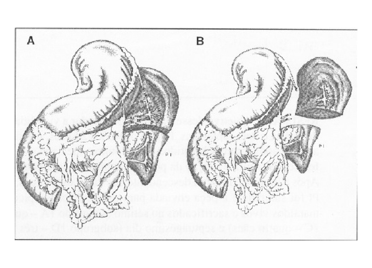
Figure 1 – Sequence of the subtotal splenectomy technique applied to dogs. A- The arrow shows the ligature and section of the vessel close to the spleen wall and the stomach, including the splenic branches. Observe in B that the inferior pole is supplied by existing vessels in gastrosplenic ligament which are can be expected to contribute to inferior lobar artery in that pole.
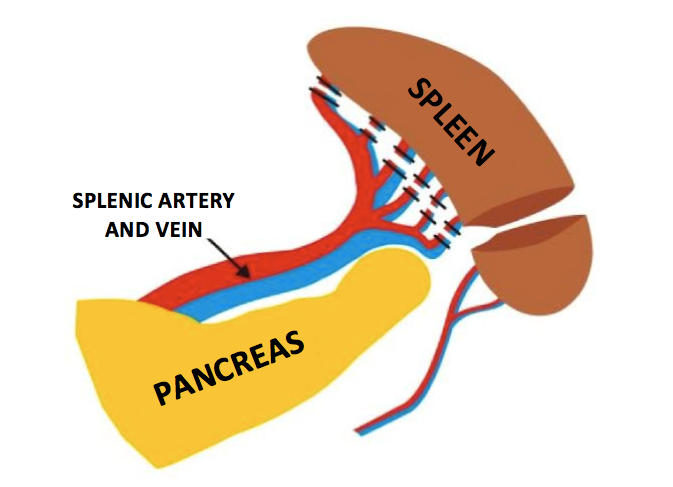
Figure 2 – ESTPI in rats. Scheme showing the vascular bandages skiming the spleen and the splenic section with upper portion removal and inferior pole maintenance (Figure – courtesy from Dr. Isabel Cristina Andreatta Lemos Paulo)
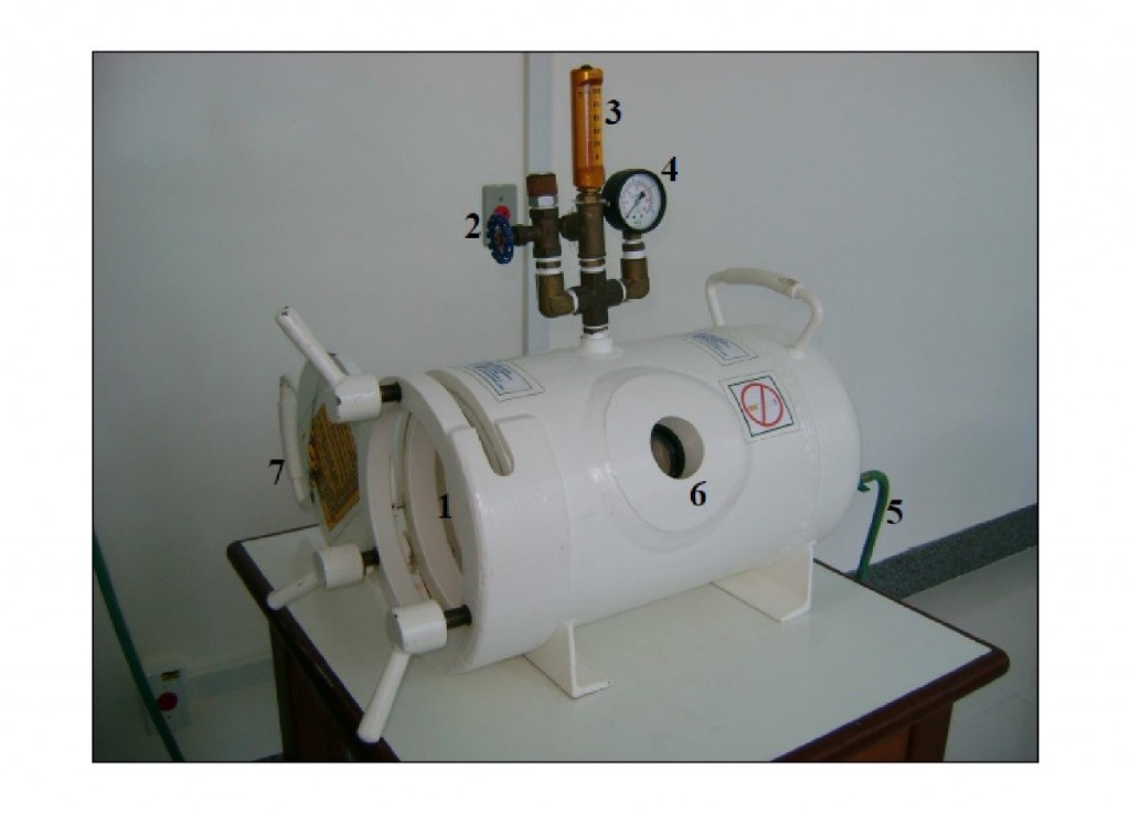
Figure 3 – Hyperbaric chamber with cylinder capsular format built with carbon steel.
1 Entry and exit the chamber; 2 Valve for oxygen escape; 3- thermometer; 4- gauge; 5- tube for oxygen inlet into the chamber; 6- display for observing the animals within the chamber; 7 – Chamber occluding cover. The chamber is 27 cm width, 51cm length and 1.5cm thick.
Authors
Danilo Nagib Salomão Paulo – Doctor/ Science School of Santa Casa de Misericórdia of Vitória (EMESCAM) (Titular Professor)
Marcela Souza Lima Paulo – Doctor/ Science School of Santa Casa de Misericórdia of Vitória (EMESCAM) (Adjunct Professor)
